Vitreo Retinal Surgery
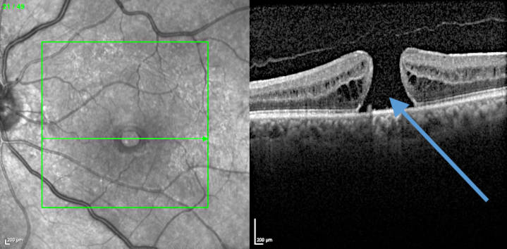
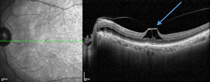
The retina is a very thin layer (about two hundred micro meters) of neural that allows the transmission of visual signals to the brain. The major retinal diseases of surgical interest are described below.
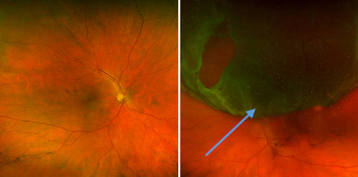
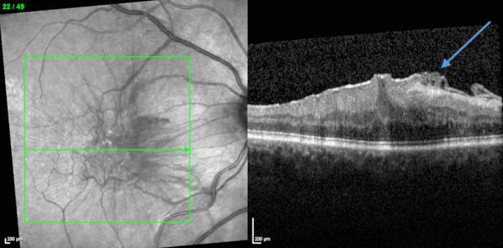
- The retinal detachment is a condition in which the neurosensory retina detaches from its normal position. It is usually associated with a peripheral retinal tear (rhegmatogenous retinal detachment). Other times it is the consequence of a pathological traction that pulls the retina away from its physiological position (tractional retinal detachment).
- The first clinical signs of retinal detachment are: flashes, flying flies, a black veil or a drop in visual acuity (when the center of the retina (the macula) is affected
- The main risk factors are age, myopia, history of ocular trauma.
- Retinal detachment needs urgent care. The surgical procedure can be carried out by an ab externo approach (the so called scleral buckling procedure) or by an ab interno one, this latter is called vitrectomy.

Vitreo retinal surgery techniques have advanced quite a lot these last few years
The anesthesia is sometimes simplified (local anesthesia), the instruments used are thinner (their caliber is 0.5 mm for 25G instruments), retinal visualisation is also much better with panoramic view systems available. With all of these combined technological advances, surgical procedures are done faster, which translates into more overall comfort for the patient.
You can make an appointment with one of the Ophthalmologists of the department who provide specific care dedicated to this pathology:

Dr Agnès Glacet-BernardStaff Physician Associated Professor at College of Medicine of Paris Hospitals

Dr Oudy SemounStaff Physician


